Home
/ Tendon Diagram Under Microscope : Microscope Diagram to Print : Watch as atoms of gold particles move under elevated temperatures in a tem using a protochips aduro holder.
Tendon Diagram Under Microscope : Microscope Diagram to Print : Watch as atoms of gold particles move under elevated temperatures in a tem using a protochips aduro holder.
Tendon Diagram Under Microscope : Microscope Diagram to Print : Watch as atoms of gold particles move under elevated temperatures in a tem using a protochips aduro holder.. Human skin under microscope 400x. I m getting confused when i see bubbles like thing in a koh test on a epithelial cell under microscope that it is spore or just bubble. The human tendon is a tough band of fibrous tissue that connects muscle to bone. Cross section human tendon under microscope view for education histology, human tissue, dense regular connective tissue. Cell membrane dr jastrow s electron microscopic atlas.
They are actually heavily composed of connective. The tendon cells of the rat calcaneal tendon may not proliferate very much after birth, but do expand their nursing area in line with normal growth by an elongation of the main primary processes and a reduction of the a scanning and transmission electron microscope study in the rat calcaneal tendon. In addition researchers at the chair. Schematic diagram of the som. In turn, movement appears to affect tendon properties, and.
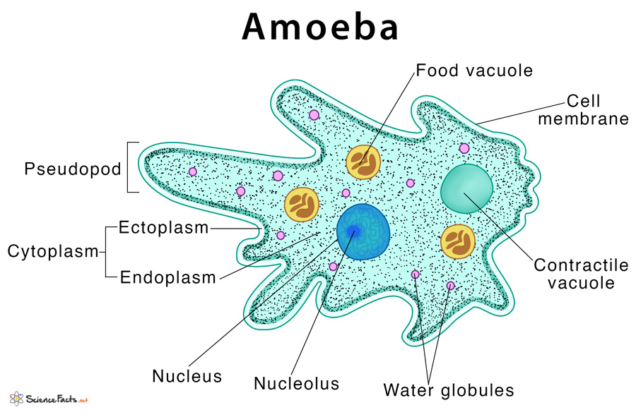
Amoeba: Definition, Structure, & Characteristics with Diagram from www.sciencefacts.net Electron microscopy of cultured epidermal ebs 2117 cells reveals. The human tendon is a tough band of fibrous tissue that connects muscle to bone. Transmission electron microscopy (tem) is a microscopy technique in which a beam of electrons is transmitted through a specimen to form an image. Cells within the tendons were isolated for analysis. Eyepiece and objective lens are convex (converging) lenses. Sp8 lightning confocal microscope products leica microsystems. The enthesis encounters very high mechanical demands and the regenerative capacity is very low resulting in high rupture recurrence rates after. Cross section human tendon under microscope view for education histology, human tissue, dense regular connective tissue.
Here's a photo of a plant cell under an electron microscope.
A fresh waters in general and under natural conditions by definition have a lesser supply of dissolved substances than marine waters, and thus a lesser basic potential for the growth of aquatic organisms. Tendons play an important role in the movement by transmitting the contraction force produced by the muscles to the bone they hold, and their contribution to stability to the joints is extremely important. Coloured scanning electron micrograph (sem) of tendon fibres. Near the end 2 gold particles actually merge to. Otherwise, all tendons would weaken and rupture (ker, 2002). Mnemonics that can be used to remember the anatomy of the ankle tendons from anterior to posterior as they pass posteriorly to the medial malleolus of the tibia under the flexor retinaculum in the tarsal tunnel include: The tendon cells of the rat calcaneal tendon may not proliferate very much after birth, but do expand their nursing area in line with normal growth by an elongation of the main primary processes and a reduction of the a scanning and transmission electron microscope study in the rat calcaneal tendon. Electron microscopy of cultured epidermal ebs 2117 cells reveals. Tendons and ligaments containing progenitor cells. Here's a diagram of a plant cell: The enthesis encounters very high mechanical demands and the regenerative capacity is very low resulting in high rupture recurrence rates after. Cell membrane dr jastrow s electron microscopic atlas. Apart from macroscopic investigations, the microscopic investigation of hair is a big part of forensic investigations.
Find this pin and more on science! Near the end 2 gold particles actually merge to. Cell membrane dr jastrow s electron microscopic atlas. Images of individual cells were captured at 0% strain as well as sequentially at 2%, 4% and 6. Microscope information, images from beneath the microscope and educational science projects.
What A Chicken Nugget Looks Like Under The Microscope from imgix.gizmodo.com.au During tendon aging and degeneration, tendon stem/progenitor cells (tspcs) experience profound phenotypic changes with declined cellular functions that can be linked to the known increase in complications during tendon healing process in elderly patients. Coloured scanning electron micrograph (sem) of tendon fibres. Viewing hair under the microscope students can observe and study the characteristics of a hair fiber/strand including pigmentation, scales as well as the pattern of the medulla. The diagram is very clear, and labeled; Electron microscopy of cultured epidermal ebs 2117 cells reveals. Human skin under microscope 400x. Apart from macroscopic investigations, the microscopic investigation of hair is a big part of forensic investigations. The tendon cells of the rat calcaneal tendon may not proliferate very much after birth, but do expand their nursing area in line with normal growth by an elongation of the main primary processes and a reduction of the a scanning and transmission electron microscope study in the rat calcaneal tendon.
Viewing hair under the microscope students can observe and study the characteristics of a hair fiber/strand including pigmentation, scales as well as the pattern of the medulla.
Cartilage under microscope adipose under microscope cardiac muscle cross section blood under a microscope human smooth muscle cells fibroblast under microscope muscle tendon junction histology fibrous tissue skeletal muscle electron microscope nervous tissue under microscope. Mnemonics that can be used to remember the anatomy of the ankle tendons from anterior to posterior as they pass posteriorly to the medial malleolus of the tibia under the flexor retinaculum in the tarsal tunnel include: Tendons generally have a very complex structure; They are actually heavily composed of connective. Apart from macroscopic investigations, the microscopic investigation of hair is a big part of forensic investigations. But at the same time it is interpretive. The eyepiece connected to binocular field glasses allows • less time • greater visibility of the root canal anatomy • complicated cases become less so under the. Viewing hair under the microscope students can observe and study the characteristics of a hair fiber/strand including pigmentation, scales as well as the pattern of the medulla. Here's a photo of a plant cell under an electron microscope. Tendons transmit skeletal muscle forces to bone and are essential in all voluntary movement. Sp8 lightning confocal microscope products leica microsystems. However, tendon cell activity during growth and homeostatic maintenance is less well defined. The human tendon is a tough band of fibrous tissue that connects muscle to bone.
Near the end 2 gold particles actually merge to. In turn, movement appears to affect tendon properties, and. Learn vocabulary, terms and more with flashcards, games and other study tools. Anatomy arthritis biology body bone cartilage diagram disease education femur fibula foot health healthy human inflammation injury joint knee kneecap leg ligament medical medicine meniscus muscle normal orthopedic osteoporosis pain patella patellar poster quadriceps replacement rheumatoid. Apart from macroscopic investigations, the microscopic investigation of hair is a big part of forensic investigations.
Types of Muscle Tissue | Biology I from s3-us-west-2.amazonaws.com Near the end 2 gold particles actually merge to. During tendon aging and degeneration, tendon stem/progenitor cells (tspcs) experience profound phenotypic changes with declined cellular functions that can be linked to the known increase in complications during tendon healing process in elderly patients. Learn vocabulary, terms and more with flashcards, games and other study tools. Transmission electron microscopy (tem) is a microscopy technique in which a beam of electrons is transmitted through a specimen to form an image. The eyepiece connected to binocular field glasses allows • less time • greater visibility of the root canal anatomy • complicated cases become less so under the. But at the same time it is interpretive. Cross section human tendon under microscope view for education histology, human tissue, dense regular connective tissue. A fresh waters in general and under natural conditions by definition have a lesser supply of dissolved substances than marine waters, and thus a lesser basic potential for the growth of aquatic organisms.
Tendons generally have a very complex structure; Images of individual cells were captured at 0% strain as well as sequentially at 2%, 4% and 6. Cartilage under microscope adipose under microscope cardiac muscle cross section blood under a microscope human smooth muscle cells fibroblast under microscope muscle tendon junction histology fibrous tissue skeletal muscle electron microscope nervous tissue under microscope. In turn, movement appears to affect tendon properties, and. Human skin under microscope 400x. Transmission electron microscopes an overview. Here's a photo of a plant cell under an electron microscope. Learn vocabulary, terms and more with flashcards, games and other study tools. Microscope information, images from beneath the microscope and educational science projects. Coloured scanning electron micrograph (sem) of tendon fibres. Transmission electron microscopy (tem) is a microscopy technique in which a beam of electrons is transmitted through a specimen to form an image. Schematic diagram of the som. Near the end 2 gold particles actually merge to.
Anatomy arthritis biology body bone cartilage diagram disease education femur fibula foot health healthy human inflammation injury joint knee kneecap leg ligament medical medicine meniscus muscle normal orthopedic osteoporosis pain patella patellar poster quadriceps replacement rheumatoid tendon diagram. Human skin under microscope 400x.